CSM Database Demo
Primary tabs
Description
The CSM database is a repository built using the Web Interfacing Repository Manager. Ported to synapse:/opt/wirm from sphenoid.
Associated project
Demo status
Runs on
Screenshots
Home
Main Menu
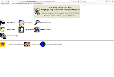
Help page
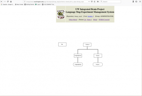
Overall Patient schema. Patients have one or more imaging studies and one or more surgeries
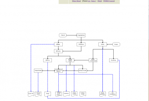
Imaging study schema. An imaging study consists of one or more MRI exams, and possibly an FMRI study. An MR Exam consists of one or more MR Series, each of which consists of a set of MR Slice. The slices can be assembled into an Aligned 3-D Volume. The volumes are used to fit a radial model containing knowledge of expected shape, which is then used to derive a surface model. A Scene consists of several surface models showing anatomy, veins and arteries. A Rendering is a 2-D snapshot of a surface model.
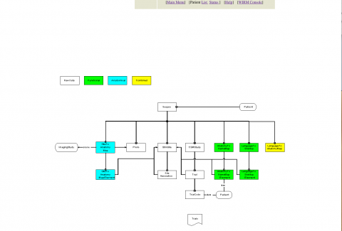
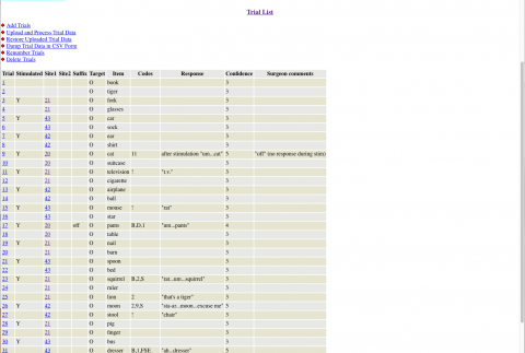
A Surgery includes one or more CSM Studies, each having a series of Trials. Small sticky numbered tags are placed on the exposed cortex and the patient is asked to name a presented object while the site is electrically stimulated. The sites are shown on an accompanying Photo. Post surgery our graphics software is used to locate the numbered sites in 3-D space in a Site To Anatomy Map, and to provide names from our Foundational Model of Anatomy (FMA) in an Anatomy To Name Map. Also post-surgery the patient responses to stimulation are coded using TrialCodes.
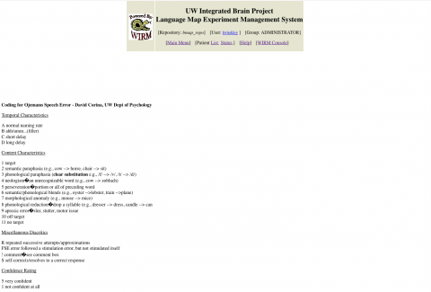
Trial codes used in analyzing a CSM study.
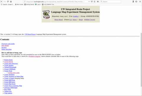
Various software tools controlled within the CSM database post surgery and imaging study allow the user to create a patient and external data directory, to create records describing the results of of the CSM Study, to Import imaging studies and 3-D Scenes, and to map sites on the photograph to 3-D surface model.

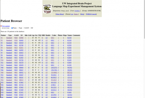
List of patients visible to an authorized user. Provides an overview of the status of the post-surgical analysis. Links take the user to more specific detail.
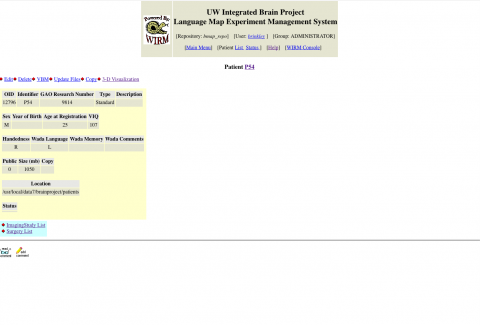
P54, a specific patient
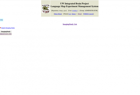
A List of imaging studies for patient P54
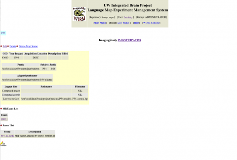
A specific imaging study, with links to MRI Exams and 3-D scenes created from MR images
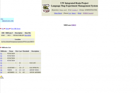
A specifc MR Exam, which consists of multiple series
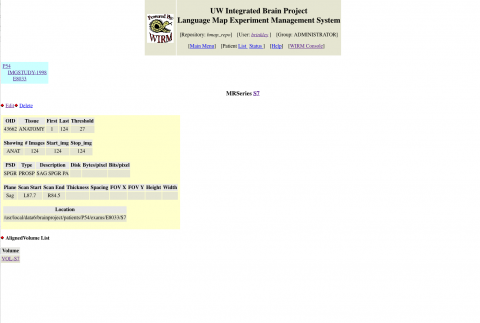
A specific MR Series optimized for anatomy, including the location of the series on our file system, and the location of a 3-D model created from slices in the series.
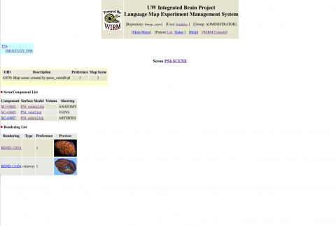
A 3-D scene combining anatomy, vein and artery 3-D models created from the relevant MR series.
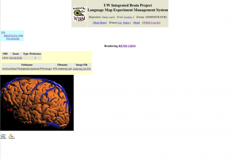
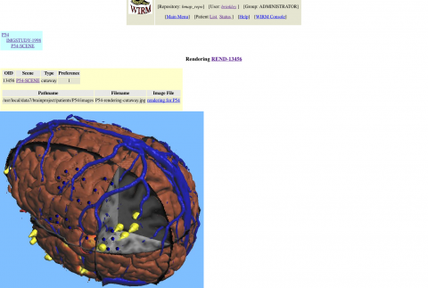
A rendering of a scene
A cutaway showing the location of sites mapped onto the 3-D scene
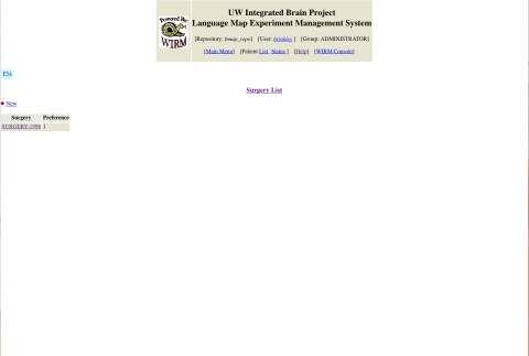
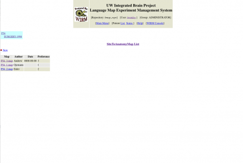
List of surgeries performed on P54 (usually only one)
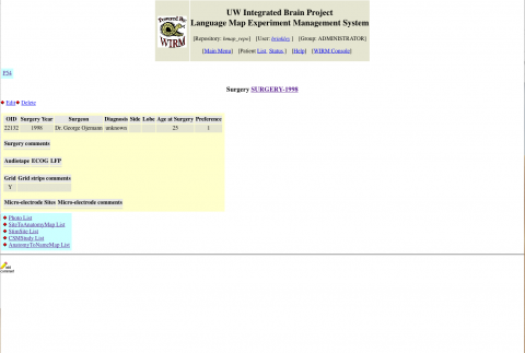
One surgery, done in 1998, plus the results of post-surgical analysis.
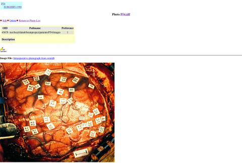
List of photos acquired during the 1998 surgery.
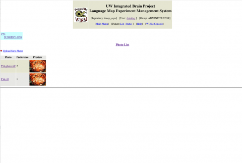
One photo, showing the tags affixed to the exposed cortical surface, where the numbers indicate stimulation sites.
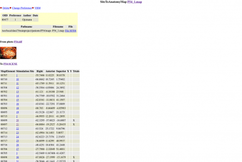
List of maps between sites on a photo and 3-D location on a 3-D scene. More than one map was often done for reproducibility.
A particular map, showing 3-D coordinates of each site within the 3-D scene.
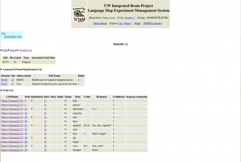
A single stimulation site, showing names of the associated anatomical structure using different parcellation schemes, and all trials done on that site.
List of CSM studies done during this surgery.
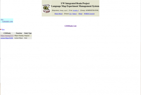
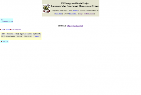
One CSM study.
CSM trials done during a single CSM study. Each trail is done on a single stinulation site, and consists of a common object shown to the patient while the site may be electically stimulated. The patient is asked to name the object. The patient response is transcribed and coded using the Corina Codes.
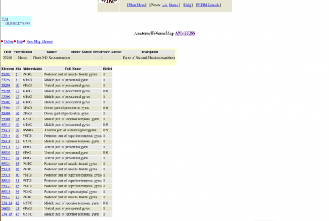
List of maps between sites and anatomcal names, using one or more parcellations and possible more than one researcher.
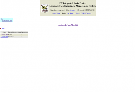
One anatomy to name map, showing the association between site numbers and anatomical names.
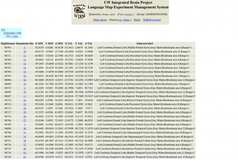
Alternate parcellation showing stimultation sites in MNI coordinates with Talairach labels